Brief About Glaucoma
Key Feature
There are three key features of Glaucoma:
Increased IOP:
The pressure inside the eye is measured with NCT (Non-Contact Tonometer) Goldmann applanation tonometer. A prism with blue lights touches the eye to accurately check IOP.
Cupping and Atrophy of the Optic Nerve:
It is the drying up of the nerve of sight as it suffers damage due to high pressure inside the eye. It is assessed by examination of the fundus of the eye.
Types of Glaucoma
- Open Angle Glaucoma
Closed Angle Galucoma
Symptoms
Unfortunately, there are no symptoms. The disease may affect 80% of the vision before a person realizes the effect on the vision. A person with chronic Glaucoma is unaware of the disease. It is the silent killer of vision. On the other hand, Acute Glaucoma, in which pressure rises rapidly, causes severe symptoms that force the patient to consult the doctor.
- Appreciation of Blind area
Poor Night Vision
- Headaches during dusk and dawn
- Pain in eyes associated with smoky vision
- Halos around light
Frequent change in number of reading glasses

Diagnosis and early detection
RISK FACTORS
People with high Intraocular Pressure have a higher risk of developing optic nerve damage. Other important risk factors include advancing age, high myopia (nearsighted), family history of Glaucoma, presence of Diabetes, past injury to the eye, surgery, or history of severe anemia or shock. We will weigh all these factors before deciding whether the patient needs treatment for Glaucoma or not. If your risk of developing Glaucoma is higher than usual but there is no damage, you will be monitored periodically as a “Glaucoma Suspect”.
High end Technology for Diagnosis
The slow death of nerve fibers is the earliest change in Glaucoma. This nerve fiber layer damage is picked by an instrument called OCT. Visual Field defects are missing as are in the field of sight, though the person may be seeing well otherwise. This is measured with an instrument called perimeter(HVF). The modern perimeter is computerized to evaluate, self-analyze, compare and report the defects.


Early Detection
A regular eye examination at WecareSecurex Clinique is the best place to detect Glaucoma. During a complete workup for Glaucoma, we will measure the Intraocular Pressure (Tonometry), the central corneal thickness (Pachymetry), inspect the drainage angle of the eye (Gonioscopy), evaluate for optic nerve head damage (Ophthalmoscopy), test the visual field of each eye (Perimetry), assess for retinal nerve fiber layer OCT.
Types Of Glaucoma
The drainage portion of the eye, called the “angle” is like a sieve and can get blocked in different ways.
Open Angle Glaucoma-It gets suddenly blocked by the iris closing off the angle. Eye pressure increases rapidly, resulting in sudden blurring of vision, severe eye pain, headache, rainbow halos around light, nausea, and vomiting. It is an emergency and if not treated immediately leads to blindness. A small hole in the iris with a laser prevents the attack of “Primary Angle Closure Glaucoma”.
Closed Angle Glaucoma- In the second type of Glaucoma, the outflow sieves get slowly blocked. This leads to an insidious rise in pressure, known as “Primary Open Angle Glaucoma”. It damages vision so gradually and painlessly that a person is unaware of the trouble until the optic nerve is badly damaged. It has no symptoms. This type of Glaucoma is much more common.

Glaucoma can also occur secondary to injury, inflammation of the eye, drugs, cataracts, etc. Glaucoma may rarely be present at birth. The parents may notice their baby’s eye enlarging and hazy(since a baby’s eye is more elastic than an adult’s). The infant or child should be taken to an ophthalmologist immediately.

Laser Treatment
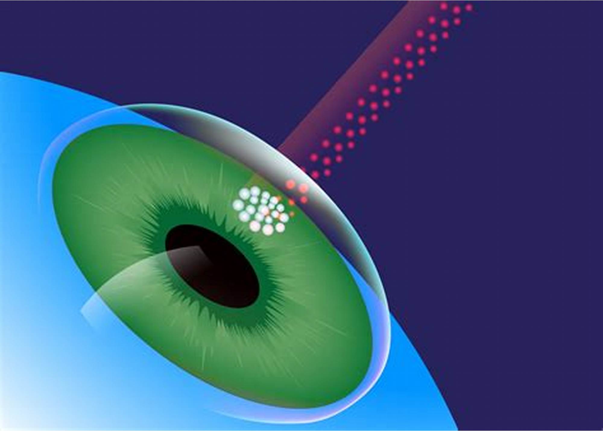
Laser surgery is effective for some types of Glaucoma. In chronic open angle Glaucoma, the drain itself is treated (trabeculoplasty), and the laser helps to reduce the medications to control the pressure. In angle-closure Glaucoma, a hole is made in the iris (Laser iridotomy) to restore the free flow of aqueous fluid.
Treatment available
Medical Treatment
Eye drops are the first line of treatment. They act to decrease eye pressure either by reducing the production of aqueous fluid within the eye or by improving the outflow through the drainage angle. Instill drops properly and regularly at prescribed timings. Tablets may be required at times. Medication should never be stopped or changed without consulting your ophthalmologist. Frequent eye examinations and tests are crucial to monitor any changes in your Glaucoma.
Surgical Treatment
Surgery for Glaucoma would be recommended only if the medicines fail to prevent damage to the optic nerve. Whatever may be the approach, the objective of the treatment is to lower the eye pressure to a level at which optic nerve damage does not develop or worsen.
In advanced cases, surgery (trabeculectomy) is necessary to control Glaucoma. Miniature instruments are used to create a new drainage channel for fluid to leave the eye, thus lowering the pressure in advanced cases. If surgery fails, special Glaucoma valves can be implanted. Fortunately, serious complications of modern Glaucoma surgery are rare.
